 english

Ostatnia modyfikacja podstrony: 27.01.2017 12:16
Project CADOCT
"Maximizing informative content of low quality OCT scans for modern computer-aided diagnostic procedures".
Motivation:
Algorithms for biometric analysis of medical images of the retina, which are the subject of this proposal, are a valuable tool for clinicians, and lead to expansion of diagnostic procedures. Implementation of non-invasive diagnostic methods, such as optical coherence tomography (OCT) in modern visualization systems allows to investigate the causes of ever more frequent lesions in the eye. Factors that deteriorate visual acuity are mainly pathogenic changes, but also natural processes in the eye, that appear at the late age.
Objective:
The main objective of this project is to develop a new method for retina image parameterization as well as an improvement of the existing approaches by combining eye features into one integrated multilevel hierarchical model.
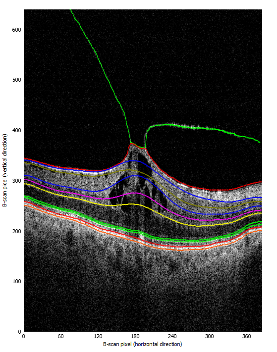
Proper detection of the retinal layers and existing pathologies is critical for proper diagnosis and further treatment.
Expected results:
This project, realized in the area of biometric image analysis, will result in the development and tests of new refined methods to detect and quantify vitreoretinal interface pathologies. The proposed solutions will contribute to scientific development of medical imaging technologies and boost the preparation of advanced diagnostic software solutions. Open-source application prepared during planned research will have help other scientists in the study of VRI pathologies and improvement of medical image analysis procedures, while OCT database will be useful to research groups worldwide investigating ophthalmic pathologies.
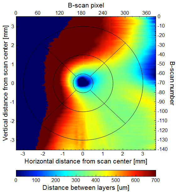
This work will provide the eye doctors community with a new tool to study VRI interface that presents data in a format intelligible for clinicians such as virtual maps of epiretinal pathologies in the macular region. These maps are meant to support diagnostic procedures, to monitor progression of the disorder and to select case-appropriate treatment algorithms. They will allow to improve the efficiency and time consumption of performed procedures. The proposed research has the potential to significantly impact the clinicians’ approach to patients with VRI pathologies.
 |
 |
Software:
Disclaimer
Please note that this project is a "work-in-progress", meaning many features still need to be implemented (see issues for the project source code). For now this application is used mainly for investigational purposes - visual analysis of retina layers depth based on existing segmentations (such as those provided by OCTExplorer application). Additionaly, for now the software is compatibly only with Avanti RTvue 3D Retina scans.
Contact:
Agnieszka Stankiewicz MSc
agnieszka.stankiewicz[at].put.poznan.pl